EndoAxis Clinical Team
Reframing the Conversation
The discussion surrounding obesity often becomes polarized: weight-centric medicine on one side, and the health-at-any-size (HAES) movement on the other. While HAES has been essential in challenging stigma, oversimplification in either direction risks obscuring the true driver of metabolic disease.
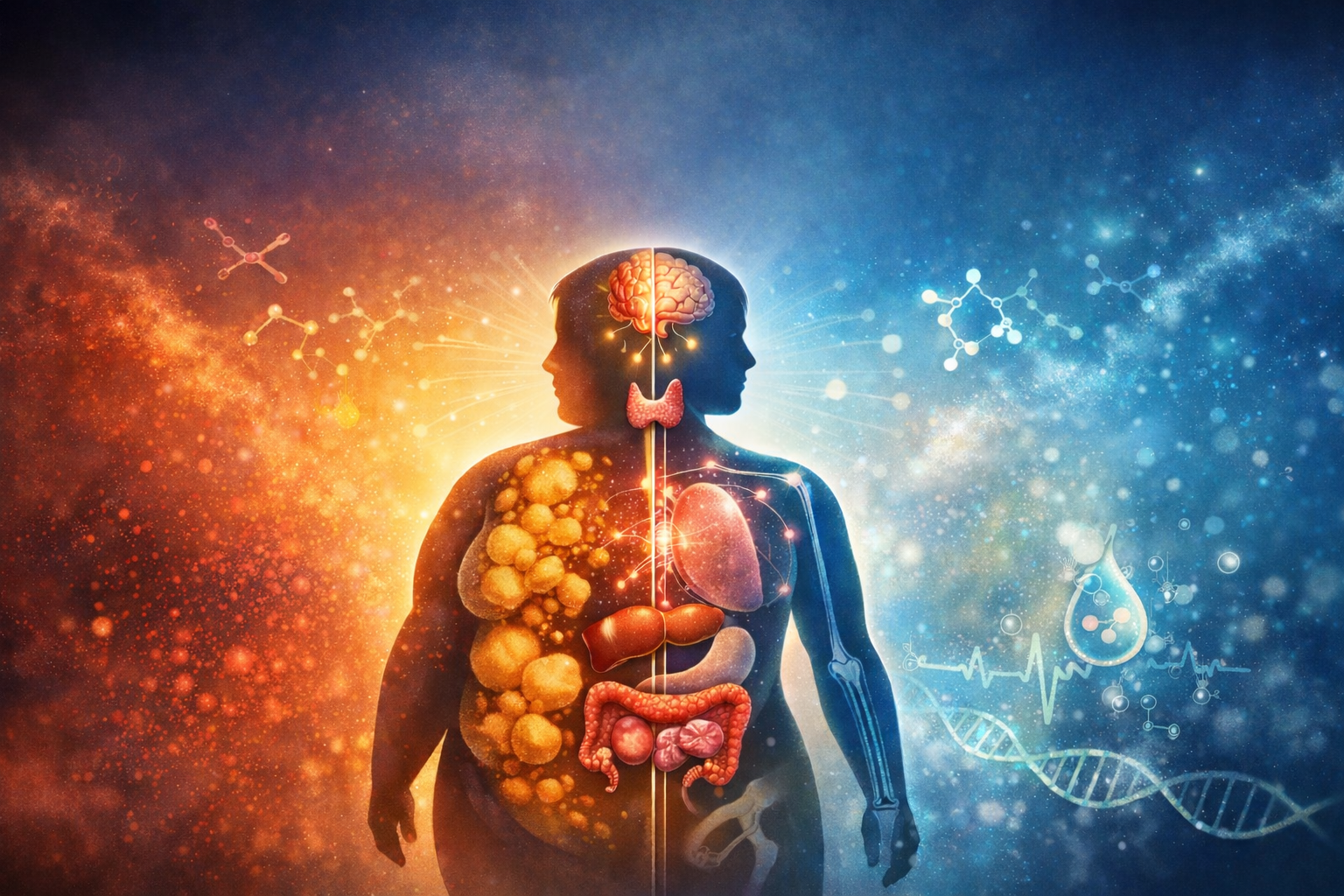
From an endocrine perspective, the issue is not body weight alone: it is adipose tissue function.
Adipose tissue is not a passive energy reservoir. It is an active endocrine and immune organ capable of both protecting and destabilizing metabolic health. Understanding when adipose tissue supports physiology — and when it disrupts it — is central to modern endocrine care.
Adipose Tissue as an Endocrine Organ
Adipose tissue actively secretes a wide range of biologically active molecules, including:
• leptin
• adiponectin
• resistin
• tumor necrosis factor–α (TNF-α)
• interleukin-6 (IL-6)
• angiotensinogen
• free fatty acids
• aromatase-derived estrogens
Through these signals, adipose tissue communicates directly with the hypothalamus, pancreas, liver, ovaries, testes, and immune system.
In a healthy state, adipose tissue functions as a buffer system, safely storing excess energy and participating in hormonal regulation. However, as adipose expands beyond its physiological capacity, its signaling profile changes, shifting from regulatory to inflammatory.
This transition, rather than body fat quantity alone, marks the beginning of metabolic dysfunction.
The Physiologic Role of Body Fat
Importantly, adipose tissue is not inherently harmful. Adequate fat mass is required for normal endocrine function, particularly in women.
Female Hormone Dependence on Adipose Tissue
In females, adipose tissue contributes meaningfully to steroid hormone dynamics. Androgens such as androstenedione are converted through peripheral pathways into testosterone and estrogens. When body fat drops below a critical threshold, typically around 16–18%, hypothalamic signaling often suppresses reproductive function.
This is not pathology, but adaptive biology: the female endocrine system interprets insufficient fat mass as an unsafe environment for reproduction.
Thus, low body fat can be as hormonally disruptive as excess adiposity.
Male Hormone Physiology
In contrast, male hormone production is less dependent on adipose tissue. Testosterone synthesis occurs primarily in the testes, allowing many men to maintain endocrine stability even at body fat levels as low as 8–10%. This sexual dimorphism explains why women experience reproductive disruption at higher body fat thresholds than men, a difference rooted in biology, not behavior.
Factors That Contribute To Bone Loss
Alcohol exposure
Alcohol disrupts bone physiology through multiple mechanisms:
- Inhibition of osteoblast differentiation
- Increased oxidative stress within bone tissue
- Altered vitamin D metabolism
- Increased cortisol secretion
- Disruption of sex hormone metabolism
Even moderate chronic intake has been associated with increased fracture risk independent of bone mineral density, underscoring alcohol’s role in impairing bone quality rather than density alone.
Medications and iatrogenic contributors
A growing number of commonly prescribed medications influence bone remodeling:
- Glucocorticoids: direct osteoblast suppression and increased osteoclast survival
- Proton pump inhibitors: impaired mineral absorption and altered osteoclast signaling
- SSRIs: serotonin-mediated inhibition of osteoblast activity
- Aromatase inhibitors and GnRH agonists: profound sex hormone depletion
- Anticonvulsants: altered vitamin D metabolism
- Chronic opioids: hypogonadism and reduced mechanical loading (physical stress)
GLP-1 receptor agonists have emerged as a newer area of concern. While not inherently osteotoxic, rapid weight loss reduces mechanical loading, often results in lean mass loss, and may reduce anabolic insulin and IGF-1 signaling to bone. In peri- and postmenopausal women, this combination may accelerate bone loss if not proactively monitored.
Endocrine and metabolic influences
Bone is an insulin-sensitive tissue. Insulin resistance, low energy availability, and chronic inflammation impair osteoblast activity and reduce remodeling efficiency.
Additional contributors include:
- Subclinical hyperthyroidism
- Elevated or dysregulated cortisol
- Protein insufficiency
- Magnesium deficiency
- Vitamin K2 insufficiency
- Zinc deficiency
- Chronic low alkaline phosphatase, suggesting impaired bone formation capacity
These metabolic stressors often coexist, compounding skeletal vulnerability over time.
The Nervous System–Bone Connection: An Underappreciated Driver
Bone is richly innervated by the sympathetic nervous system. Norepinephrine acts on β2-adrenergic receptors expressed on osteoblasts, suppressing bone formation and favoring resorption.
Chronic sympathetic activation, whether from psychological stress, sleep disruption, metabolic disease, or circadian misalignment, shifts bone remodeling toward loss. This neuro-skeletal axis reframes osteoporosis as partially a disorder of autonomic signaling rather than purely mineral deficiency.
Aromatase Activity and Hormone Balance
Adipose tissue expresses aromatase, the enzyme responsible for converting androgens into estrogens.
In physiologic amounts, this conversion is protective and necessary. However, with excess adipose mass, aromatase activity increases substantially.
In Women
Increased aromatization may lead to:
• elevated estrone and estradiol production
• relative progesterone deficiency
• functional estrogen dominance
• worsened insulin resistance
• increased risk for estrogen-sensitive conditions
Notably, higher estrogen production does not equate to improved estrogen signaling. Excess or poorly cleared estrogens may amplify inflammatory and metabolic burden rather than benefit.
In Men
In males, increased aromatase activity converts testosterone into estradiol, contributing to:
• reduced free testosterone
• suppression of LH and FSH
• decreased lean mass
• increased fat accumulation (belly, man boobs)
This creates a self-reinforcing endocrine loop in which rising adiposity further suppresses androgen signaling.
When Adipose Tissue Becomes Pathologic
Adipose tissue becomes metabolically harmful not simply when it expands, but when it becomes dysfunctional.
Characteristics of unhealthy adipose tissue include:
• adipocyte hypertrophy
• tissue hypoxia
• macrophage infiltration
• chronic low-grade inflammation
• insulin resistance within fat cells
At this stage, adipose tissue loses its capacity to safely store triglycerides. When lipids spill into circulation, and subsequently into tissues like the liver, muscle and pancreas, it accelerates insulin resistance and metabolic disease.
In this context, adipose tissue behaves less like storage and more like a source of endocrine stress signaling.
Health at Any Size: Where the Model Holds — and Where It Does Not
The HAES framework has contributed meaningfully to clinical care by emphasizing:
- Reduced weight stigma
- Improved patient engagement
- Focus on behaviors over scale weight
- Recognition that BMI alone is not diagnostic
These principles are well supported.
However, HAES has physiological limits.
Health at any size speaks to human worth, not endocrine neutrality.
Two individuals with identical BMI values may exhibit dramatically different metabolic profiles:
- One with preserved insulin sensitivity, adequate muscle mass, and low inflammatory burden (best case)
- Another with visceral adiposity, hyperinsulinemia, fatty liver, and impaired mitochondrial function (worst case)
Similarly, individuals with normal or low body weight may carry significant metabolic risk, often referred to as “normal-weight obesity” or “skinny fat” physiology.
Weight alone does not define health, but adipose dysfunction does.
The Central Issue: Inflamed Fat
The unifying driver across obesity-related disease is inflamed adipose tissue.
When fat becomes insulin resistant and inflammatory, it contributes to:
- Hyperinsulinemia
- Suppressed sex hormone–binding globulin (SHBG)
- Worsened ovarian and adrenal androgen imbalance
- Estrogen dysregulation
- Increased cardiometabolic risk
Thus, the clinical goal is not leanness, but adipose resilience.
Healthy fat tissue can support endocrine function. Inflamed fat tissue cannot, no matter the size of a person.
Clinical Implications
From a practitioner standpoint, the focus should shift away from weight-centric targets and more toward restoring adipose health through:
- improving insulin sensitivity
- Reducing inflammatory burden
- Supporting circadian rhythm and sleep
- Increasing skeletal muscle mass
- Optimizing hormonal clearance and signaling
When adipose tissue becomes metabolically flexible again, changes in weight, if they occur, follow as a downstream effect rather than a primary objective.
Conclusion
Adipose tissue is neither villain nor virtue. It is an endocrine organ whose function determines metabolic outcome.
The goal of care is not to eliminate fat, nor to ignore its biology, but to restore its capacity to communicate appropriately within the endocrine system.
Health is not defined by size alone, but neither is size irrelevant when adipose tissue becomes inflamed, dysregulated, and hormonally disruptive.
Understanding this distinction allows clinicians to move beyond ideology and toward physiology, where sustainable metabolic healing truly begins.
Stay tuned for next week, when we will dive into urinary metabolite patterns that may indicate when adipose has become dysfunctional, and how our EndoAxis technology and products may support these underlying factors!
In Health,
The EndoAxis Team